Efficient retina examination is essential for accurate ophthalmic diagnosis and timely patient care. As retinal disorders continue to increase due to aging, diabetes, vascular conditions, and lifestyle-related health issues, eye care professionals require advanced imaging technologies that can support faster and more reliable retinal evaluation. Modern ophthalmic diagnostics now rely heavily on detailed retinal imaging systems that improve visualization, simplify documentation, and strengthen long-term patient monitoring.
Retinal abnormalities often begin silently and may not produce symptoms during the early stages. Because of this, clinicians need precise imaging systems capable of identifying subtle structural and vascular changes before severe visual complications occur. Modern retinal imaging technologies have transformed clinical workflows by enabling detailed retinal analysis while reducing examination time and improving diagnostic consistency.
Advanced imaging systems also support better communication between specialists, improved treatment planning, and enhanced preventive eye care strategies. These advantages make modern retinal imaging an essential part of efficient ophthalmic practice.
Importance of Efficient Retina Examination
Retina examination plays a critical role in identifying diseases that affect visual function and retinal health. Since many retinal disorders progress gradually, timely and accurate evaluation is extremely important for preserving long-term vision.
Efficient retinal examination supports:
- Early disease detection
- Faster clinical assessment
- Improved treatment planning
- Better patient management
- Reduced risk of delayed diagnosis
As patient volume increases in ophthalmic clinics, efficient retinal imaging becomes increasingly valuable for maintaining both diagnostic quality and workflow performance.
Challenges in Traditional Retinal Evaluation
Traditional retinal examination methods may sometimes require extended examination time or may not provide detailed documentation for long-term monitoring. Subtle abnormalities can also be difficult to identify without high-resolution retinal imaging support.
Common challenges include:
- Difficulty visualizing microscopic retinal changes
- Limited long-term documentation
- Inconsistent comparison during follow-up visits
- Increased examination time in complex cases
Advanced retinal imaging technologies help overcome these limitations by providing detailed retinal visualization and reliable image storage capabilities.
Role of Modern Retinal Imaging Technology
Modern retinal imaging systems use advanced optical technology and controlled illumination to capture highly detailed images of retinal structures. These systems allow clinicians to evaluate retinal tissues, blood vessels, and optic nerve regions with improved clarity and efficiency.
Retinal imaging supports assessment of:
- Retinal tissue integrity
- Vascular structure
- Macular condition
- Optic nerve appearance
- Peripheral retinal regions
Detailed visualization significantly improves diagnostic confidence and supports more efficient ophthalmic evaluation.
Improving Examination Workflow Efficiency
One of the major advantages of advanced retinal imaging systems is their ability to improve workflow efficiency in ophthalmic clinics. High-resolution retinal images can be captured quickly while maintaining excellent image quality and diagnostic precision.
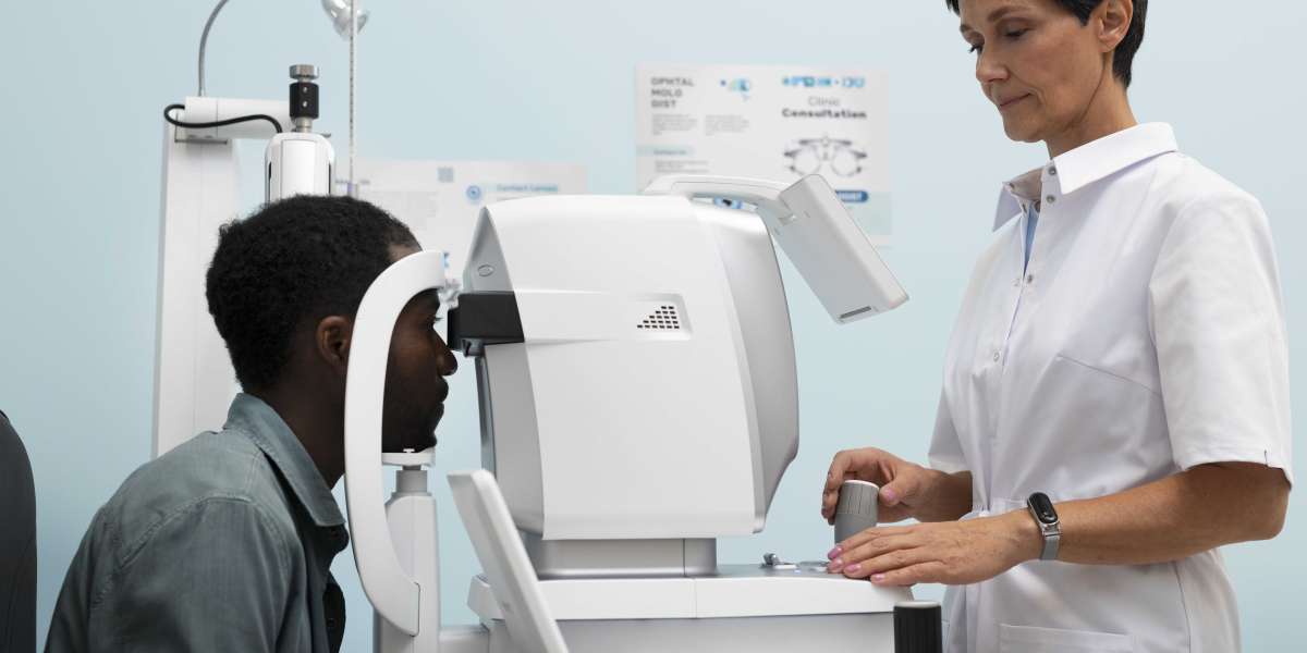
In clinical ophthalmic practice, the fundus camera helps clinicians perform efficient retinal assessments by enabling rapid image capture, detailed retinal documentation, accurate visualization of retinal abnormalities, and streamlined patient evaluation procedures.
Modern systems with automated alignment and imaging features further reduce examination complexity and support faster patient throughput.
Supporting Early Detection of Retinal Disorders
Retinal disorders often begin with subtle structural or vascular abnormalities that may remain hidden during routine examination procedures. Advanced retinal imaging allows clinicians to identify these abnormalities earlier and more accurately.
Early retinal findings may include:
- Small hemorrhages
- Retinal swelling
- Vascular irregularities
- Pigment abnormalities
- Optic nerve changes
Early detection improves treatment opportunities and helps reduce the risk of progressive visual impairment.
Enhancing Diagnostic Accuracy
High-quality retinal imaging improves diagnostic precision by providing clinicians with clear and detailed visualization of retinal structures. Accurate retinal assessment helps specialists evaluate disease severity and monitor progression more effectively.
Improved diagnostic accuracy supports:
- Better treatment planning
- Reliable disease classification
- More effective patient communication
- Consistent clinical documentation
Detailed retinal imaging also strengthens long-term ophthalmic monitoring programs.
Importance of Long-Term Retinal Monitoring
Many retinal conditions require continuous monitoring because abnormalities may progress gradually over time. Retinal imaging allows clinicians to compare images from multiple visits and identify subtle changes more consistently.
Long-term monitoring supports:
- Assessment of retinal stability
- Evaluation of treatment response
- Detection of disease progression
- Improved clinical management strategies
Maintaining detailed retinal records improves continuity of care and supports better ophthalmic decision-making.
Contribution of Advanced Ophthalmic Equipment
The effectiveness of retinal examination depends heavily on the quality of imaging systems used in clinical environments. High-performance retinal imaging equipment provides clearer visualization, enhanced contrast, and more reliable diagnostic performance.
Matronix Optotechnik provides advanced ophthalmic imaging solutions designed to support efficient retinal evaluation and streamlined clinical workflows. Their retinal imaging systems are developed with modern optical technology that enables clinicians to achieve accurate retinal visualization, faster image acquisition, and improved diagnostic confidence during ophthalmic examination procedures.
Supporting Better Clinical Documentation
Detailed retinal documentation is essential for accurate ophthalmic diagnosis and long-term patient care. High-resolution retinal images provide objective visual records that can be stored, reviewed, and compared over time.
Reliable documentation supports:
- Better collaboration between specialists
- Improved follow-up evaluation
- Accurate patient records
- More effective treatment planning
Digital retinal imaging has become a valuable tool in maintaining consistent and organized clinical documentation systems.
Advancements in Retinal Imaging Technology
Technological innovation continues improving retinal imaging capabilities and examination efficiency. Modern retinal imaging systems now include automated image capture, wide-field visualization, enhanced contrast imaging, and digital image analysis features.
Research in retinal imaging technology is also improving image quality and expanding field-of-view capabilities for better retinal assessment.
Future developments are expected to improve:
- Imaging speed
- Diagnostic precision
- Automated retinal analysis
- Preventive ophthalmic care strategies
These innovations will continue strengthening retina examination efficiency and improving patient outcomes.
Conclusion
Efficient retina examination is essential for accurate diagnosis, preventive eye care, and long-term visual health management. Advanced retinal imaging technologies have transformed ophthalmic practice by enabling detailed retinal visualization, faster image acquisition, and improved clinical workflow efficiency.
With reliable imaging systems and continued technological advancement, clinicians can improve diagnostic precision, strengthen long-term retinal monitoring, and provide better visual care outcomes for patients undergoing retinal evaluation.